Fetal care during Pregnancy
How many scans are needed for all pregnant ladies and why?
Usually there are two scans that are compulsory for every pregnant woman.
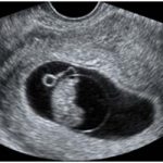
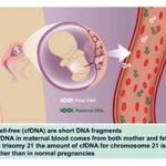
The first one is NT Scan done around 11-14 weeks. This scan is useful to demonstrate that : the pregnancy is healthy one (live fetus, in right location), to diagnose multiple pregnancies 2,3 or even more (and it is not rare to have surprises at times !), to measure the size of the fetus and thereby to calculate the expected date of delivery, to diagnose major fetal abnormalities (upto 70% of the fetal abnormalities are detected at this stage) , to screen for Downs Syndrome and major chromosomal abnormalities by measuring fluid behind fetal neck and to examine fetal nose and blood flow through fetal heart and liver. By combining the ultrasound findings with measurement of two hormones produced by the placenta we can achieve close to 95% detection rate of Down’s syndrome (combined screening).
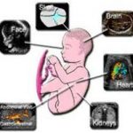
The second scan is Fetal Anomaly Scan or Morphology Scan done around 20 weeks of pregnancy. It is a detailed study of fetus (head to toe examination) to exclude any physical abnormalities in the baby (upto the extent of more than 95% of physical abnormalities can be identified). Basic study of Fetal heart is also done with anomaly scan and it is sufficient in all low risk pregnant women. However, in few situations detailed study of fetal heart is needed (known as Fetal ECHO).
The third scan is optional (better if it is possible to have it) and is done around 34-36 weeks to check the baby’s growth, Doppler test and well-being of the baby (to check baby’s weight, fluid surrounding baby and blood flow studies). It is very helpful in high risk cases to decide the timing of delivery.
Frequent scans may be required for patients with special cases like when pregnant mother has high BP (hypertension) Diabetes, Kidney Problems, Multiple pregnancies, small baby, previous bad outcomes and so on. Especially Diabetes (which is common among Asian population) can lead to fetal abnormalities like spinal and heart problem in the baby, big babies and unexplained still birth after 38 weeks.
What are the causes for fetal abnormalities (anomalies)?
✓ Fortunately,nearly 98% of the times the fetus is normal.
✓ Fetal anomalies can occur in upto 1-2% of pregnancies and unfortunately they are increasing.
✓ Reasons for fetal anomalies could be multi factorial such as environmental, consanguineous (blood relation) marriage, maternal diseases such as diabetes,some medications,infection, environmental pollution, adulteration or in vast majority of cases unknown. Fetal abnormalities may be of different types- lethal or non-lethal. Sometimes there may be insignificant findings (Soft markers or minor markers) which does not have any implications on the fetus if it is isolated. Therefore patients can be reassured here.
How Invasive testing is helpful during pregnancy?
This helps in assessing whether the fetus has any serious health condition in the form of chromosomal abnormality or few genetic abnormalities (not all the genetic abnormalities are detected by doing amniocentesis).
Some of the common indications for this test are- couple who already have had a baby with certain problems, family history of genetic problems including some rare inherited diseases, Abnormal findings on Scan, screen positive result from the Down’s Syndrome Screening etc.
There are two types of methods of Invasive Testing.
The First one is CVS(Chorionic Villous Sampling) where a small sample of tissue is taken from the placenta and is done between 11-15 weeks.
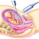
The second one is Amniocentesis (done after 15 weeks of pregnancy) where the sample of fluid surrounding the baby is taken for chromosomal studies. Both these tests are done under the ultrasound guidance using the fine needle and are helpful to find out fetal abnormalities.
What is the significance of 3D/4D scans?
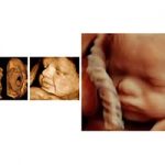
3D/4D imaging is the latest advancement in fetal ultrasound technology. 3D is still pictures and provides three dimensional view of the fetus. This scan is best done around 28 weeks of pregnancy. With very high qualities of images, it is possible to clearly see baby’s mouth, lips, nose, eyes and facial expressions. This scan is very useful for the detailed assessment of few fetal anomalies (brain, face, spine, limb problems etc.). 4D scans are moving 3D images of the baby and produces life like images so that you can actually see how the baby looks like (also known as baby bonding scans).
What are the fetal treatment procedures currently available in medical science?
In some cases, the abnormalities are more frequently recognized prior to the time of delivery. It is possible to prevent complications substantially through the methods that are accepted as standard practice.
These procedures are:
✓ Selective fetal reduction:
In cases of multiple pregnancies the fetal numbers can be reduced to desired number to reduce pregnancy complications mainly risks of abortion, extreme premature delivery etc (for eg: from quadruplets to twins ,triplets to twins).
✓ Foetal blood transfusions:
Intrauterine foetal blood transfusions to treat foetalanaemia due to various causes (commonest being mother’s blood group is negative with previous sensitisation)
✓ Laser treatment:Laser treatment of connecting blood vessels under foetoscopic (camera passed inside the uterus ! like laparoscopic surgery) guidance effective in treating twin-twin transfusion syndrome (a complication in identical twins where one baby grows bigger than other).
✓ Foetal surgeries:
Intrauterinefoetal surgeries like excision of tumors may sometimes needed to improve the outcome.
✓ Foetal shunting:
Sometimes shunting procedure needs to be done to treat fluid collection in lungs and other organs.

